
Quick Answer: After you cast an agarose gel from agarose powder (CAS 9012-36-6) and run DNA, you need a stain + imaging method to see bands. Ethidium bromide (EtBr) is the historic default (high sensitivity, UV imaging, strict waste rules). SYBR-family dyes (e.g., SYBR Safe, SYBR Green) are common lower-toxicity alternatives, often imaged with blue light. GelRed / GelGreen are another fluorescent class marketed for safer handling and blue-light compatibility. Pick the stain that matches your institutional EHS rules, imager hardware, and validated SOP — then document waste segregation for gel slices, running buffer, and stain stock.
Why Stain Choice Matters in Agarose Workflows
Casting and running the gel is only half the workflow. Weak or mismatched staining shows up as “no bands,” “faint ladder,” or “we changed agarose and nothing improved” — when the real issue is stain concentration, post-run handling, or imaging settings.
If you are still standardizing the gel itself, start with Agarose (CAS 9012-36-6): What It Is, How Gel Electrophoresis Works & How to Choose Powder. This article covers the post-run layer: stain selection, imaging, and waste disposal for Agarose (CAS 9012-36-6) gels.
EtBr, SYBR, and GelRed at a Glance
| Stain class | Typical use | Imaging light | Lab perception |
|---|---|---|---|
| Ethidium bromide (EtBr) | DNA intercalating stain — classic agarose gels | UV (302/365 nm) | High sensitivity; mutagen concerns; strict waste |
| SYBR Safe / SYBR Green I | Fluorescent DNA dyes for gels | Often blue light (product-dependent) | Lower acute toxicity vs EtBr in many SOPs |
| GelRed / GelGreen | Fluorescent DNA stains (commercial blends) | Blue light or UV (check product sheet) | Common “safer stain” alternative in teaching cores |
| Pre-stain in buffer (some workflows) | Stain mixed into gel or running buffer | Depends on dye | Convenient but can affect migration — validate SOP |
Product names vary by vendor. Always read the manufacturer SDS, imaging filter recommendations, and your institution’s chemical approval list before switching stains.
EtBr vs SYBR vs GelRed: Side-by-Side Comparison
| Topic | EtBr | SYBR-family | GelRed / GelGreen |
|---|---|---|---|
| Sensitivity (typical lab view) | Very good for routine DNA bands | Good — often sufficient for PCR checks | Good — vendor-dependent |
| Imaging hardware | UV transilluminator / gel doc | Blue-light box or compatible gel doc | Often blue light; some UV workflows |
| EHS / approval burden | Higher — mutagen labeling common | Often lower — still treat as chemical waste | Often positioned as safer — still regulated |
| Staining mode | Post-stain bath or low conc. in gel | Post-stain or in-gel (product-specific) | Post-stain typical; follow kit |
| Background / gel clarity | Can show fluorescence in gel matrix | Generally clean on agarose | Generally clean; check lot validation |
| Teaching-lab trend | Phasing out in some campuses | Common replacement path | Common replacement path |
| Downstream enzyme risk | Carryover possible — plan cleanup | Generally lower carryover concern | Follow vendor notes for recovery SOPs |
When to Choose Each Stain
Choose ethidium bromide (EtBr) when:
- Your validated SOP and regulatory filings already specify EtBr
- You have UV imagers and trained waste streams in place
- You need a historical reference method for method transfer or publication comparability
- Institutional approval still allows EtBr under controlled access
Even where EtBr remains permitted, document PPE, designated areas, and waste segregation in the SOP — not only “how to stain.”
Choose SYBR-family dyes when:
- Your campus pushes lower-toxicity DNA stains
- You image with blue-light boxes to reduce UV exposure
- Teaching labs want a stain that students recognize across kits
- You need reasonable sensitivity without EtBr waste complexity
Confirm whether your product is SYBR Safe (often marketed for gel work) vs SYBR Green I (broader use) — concentrations and imaging filters differ.
Choose GelRed / GelGreen when:
- Your core facility stocks GelRed as the default safer stain
- Vendor kits pair GelRed with specific buffer and imaging instructions
- You want an alternative fluorescent stain without returning to EtBr
- Blue-light imaging is the standard in your lab layout
Staining Workflow Options on Agarose Gels
| Method | How it works | Buyer / SOP note |
|---|---|---|
| Post-stain (after run) | Run gel → stain bath → rinse → image | Most common teaching workflow; easier waste segregation |
| In-gel / pre-cast stain | Dye in gel matrix or running buffer | Validate band migration and sensitivity vs post-stain |
| Destain / rinse step | Reduce background fluorescence | Time and buffer volume affect faint bands |
| Gel slice for recovery | Image → excise band under light | Use stain compatible with downstream enzymatic steps |
Recovery workflows may require LMP agarose — see Standard vs LMP Agarose (CAS 9012-36-6 vs 39346-81-1). Stain choice is independent of agarose grade but must appear on the same SOP.
Imaging Setup: UV vs Blue Light
Imaging is not “open the app and click.” Lock these parameters in the SOP:
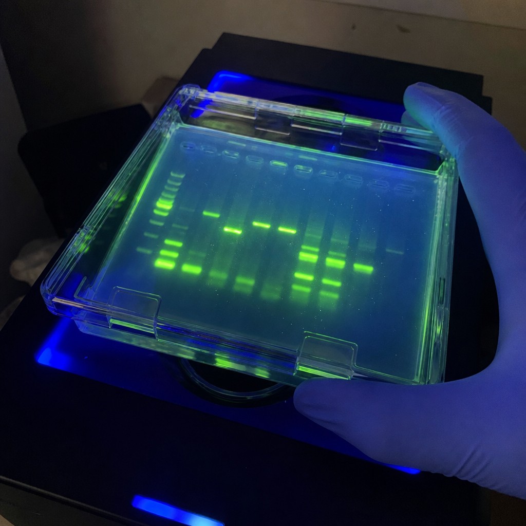
- Excitation source: UV vs blue LED — must match dye
- Exposure time: auto vs fixed — affects quantitation
- Filter / emission channel on digital gel docs
- Saturation control — saturated bands cannot be quantified
- Ladder imaged on every gel — stain performance drifts with old stock
Switching from EtBr (UV) to SYBR/GelRed (blue light) often requires a hardware check, not only a reagent swap.
Waste Disposal: What Labs Must Segregate
Gel electrophoresis generates multiple waste streams. Treat stain choice as an EHS decision, not only an imaging decision.
| Waste stream | Typical contents | Handling principle |
|---|---|---|
| Gel slices / cast gels | Agarose + DNA + stain | Segregate per institutional chemical-waste rules |
| Post-stain bath / rinse buffer | Dye + buffer salts | Often hazardous chemical waste — do not pour to sink |
| Running buffer (TAE/TBE) | Buffer + optional dye carryover | Some labs drain separately from stain waste — follow EHS chart |
| EtBr stock solutions | Concentrated mutagen (where used) | Often dedicated container; never mix with general trash |
| Gloves / wipes | Trace dye contamination | Lab trash vs chemical waste — campus rules vary |
Do not assume “safer stain” means “sink disposal OK.” SYBR and GelRed workflows still produce dye-containing liquid waste that most institutions classify as chemical waste.
Practical Waste Workflow for Core Facilities
- Label containers at the bench: gels, liquid stain waste, buffer waste (if split)
- Train users on no sink disposal for stain baths unless EHS explicitly allows
- Keep SDS for each stain at the station
- Log stain lot when bands look weak — expired dye mimics “bad agarose”
- Coordinate with building EHS for pickup schedules — full containers should not sit open
Document waste rules beside the gel station, not only in a binder — electrophoresis areas see high user turnover.
Decision Guide for Lab Managers
| Situation | Stain direction | Also lock in SOP |
|---|---|---|
| New teaching lab setup | SYBR Safe or GelRed + blue light | Waste containers + imager filters |
| Legacy UV gel doc only | EtBr (if still approved) or dye with UV option | PPE + UV shielding |
| Gel extraction for cloning | Stain with documented recovery compatibility | LMP agarose grade if required |
| High-throughput PCR checks | Post-stain SYBR/GelRed for speed | Standardize rinse time |
| Regulated QC lab | No silent stain switches | Change-control on stain lot and imager settings |
Buffer system affects run quality before staining — see TAE vs TBE for agarose gels. Gel type context: Agarose vs PAGE.
Common Staining Mistakes (and Fixes)
| Symptom | Likely cause | First fix |
|---|---|---|
| No bands visible | Wrong imager channel / exposure | Re-image with ladder; check light source |
| Very faint ladder | Expired stain or short stain time | Fresh working solution; validate timing |
| High background | Insufficient rinse | Extend rinse; reduce stain concentration |
| Smear after staining | Gel handled while warm / overloaded sample | Cool gel; reduce load; check dye in buffer |
| Bands differ from last week | Silent stain or buffer switch | Freeze stain + buffer + agarose CAS on SOP |
For broader electrophoresis troubleshooting, see nucleic acid electrophoresis troubleshooting.
What to Put on the SOP and RFQ
- CAS 9012-36-6 (or LMP CAS if recovery SOP requires)
- Gel %, buffer (TAE/TBE), voltage/time
- Stain name + vendor catalog number
- Stain mode: post-stain vs in-gel; concentration; time; rinse steps
- Imager: UV vs blue light; exposure guidelines
- Waste container labels and prohibited disposal routes
Evaluating agarose powder while you standardize staining? Request a 5–20 g sample + COA for Agarose CAS 9012-36-6 via Request a Quote or [email protected].
FAQ
Is EtBr still used for agarose gels?
Yes in some labs with legacy SOPs and UV imagers, but many institutions have moved to SYBR or GelRed for lower hazard workflows. Follow your campus chemical approval list.
Is SYBR Safe the same as SYBR Green?
Not always. They are related fluorescent dye families but differ in formulation, concentration, and imaging guidance. Use the vendor sheet for the exact product on your bench.
Can I use GelRed with UV transilluminators?
Some GelRed products can be imaged with UV as well as blue light — check the product documentation. Do not assume all “safe stains” share the same imaging protocol.
Can stain waste go down the sink?
In most academic and industrial labs, no — dye-containing liquids are collected as chemical waste. Confirm with your institutional EHS office.
Does stain choice affect which agarose I should buy?
Indirectly. Stain and imager are separate from agarose grade, but the full method (agarose CAS, buffer, stain, waste) must be validated together. Most routine work still uses standard agarose CAS 9012-36-6.
Why do my bands look fine but quantitation is wrong?
Saturated pixels, uneven illumination, and stain pooling in the gel matrix distort intensity. Use non-saturated exposures and consistent ladder loads.
Where can I source electrophoresis agarose (CAS 9012-36-6)?
Monuo Chemical supplies standard Agarose (CAS 9012-36-6) and low gelling temperature agarose (CAS 39346-81-1). Contact: Request a Quote · [email protected].
Conclusion
Pick EtBr only where still approved and paired with UV imaging and strict waste control. Choose SYBR or GelRed when your facility targets lower-toxicity stains and blue-light workflows. Whichever you standardize, write stain, imager, and waste streams into the same SOP as agarose CAS 9012-36-6, gel %, and buffer — weak bands are often a staining or imaging problem, not a powder problem.
Request a 5–20 g sample + COA of Agarose (CAS 9012-36-6) while you lock staining standards via the contact page.
Related Resources
- Agarose (CAS 9012-36-6) product page
- Agarose Low Gelling Temperature (CAS 39346-81-1) product page
- Agarose guide: electrophoresis and powder selection
- TAE vs TBE for agarose gels
- Agarose vs PAGE for nucleic acids
- Standard vs LMP Agarose comparison
- Nucleic acid gel electrophoresis overview
- Nucleic acid electrophoresis troubleshooting
- Agarose essential polysaccharide overview
- Request a Quote / Contact Monuo Chemical
- Monuo Chemical News
Disclaimer: Information is for laboratory method selection, safety awareness, and purchasing reference. Stain hazards, disposal routes, and PPE requirements vary by jurisdiction and institution — always follow your official EHS SOP and SDS. Protocols are illustrative and must be validated locally. Verify agarose lots against the supplied COA and SDS.