
Basic Principles of Separating Nucleic Acids by Gel Electrophoresis
Gel electrophoresis is a common experimental technique in molecular biology for identifying, quantifying, and purifying nucleic acids. Owing to its speed and versatility, it is widely used for the separation and analysis of nucleic acids. Using gel electrophoresis, nucleic acids ranging from approximately 100 bp to 25 kb can be separated within minutes to several hours, and high-purity nucleic acids can be efficiently recovered from the gel—most often using an agarose gel matrix (CAS 9012-36-6).
This technique applies an electric field to a mixture of charged molecules and induces migration based primarily on size, charge, and structure. In the neutral-to-alkaline pH range (Figure 1A), the phosphate groups on the nucleotide backbone of nucleic acids carry a negative charge. Therefore, each nucleotide carries a net negative charge—that is, the total charge of a nucleic acid molecule is proportional to the total number of nucleotides, or its mass.
In other words, the charge-to-mass ratio of a DNA or RNA molecule is constant. Thus, their mobility in gel electrophoresis depends mainly on differences in molecular size (when structures are comparable). Under an electric field, nucleic acids migrate from the negative electrode (cathode) toward the positive electrode (anode); shorter fragments move faster than longer ones, resulting in separation based on size (distance) (Figure 1B).
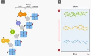
Figure 1. (A) Net negative charge carried by a nucleic acid strand. (B) Separation of nucleic acid fragments of different lengths by gel electrophoresis.
Most life-science researchers will “run a gel” at some stage of their careers. To run a gel, an electric field is applied across a matrix through which biomolecules (such as nucleic acids and proteins) can be separated according to differences in mobility, which depend on their relative size, charge, and structure. A typical gel matrix acts as a molecular sieve that impedes migration driven by the electric field. This process is called gel electrophoresis—literally, being carried by electric current through a gel. How does nucleic acid gel electrophoresis work, and how did the related techniques arise? Below we briefly outline the history of nucleic acid gel electrophoresis.
Furthermore, the migration distance of nucleic acids in gel electrophoresis usually has a predictable relationship with their size, enabling calculation of nucleic acid size in a given sample. For linear double-stranded DNA fragments, within a certain range, migration distance is inversely proportional to the logarithm of molecular weight (Figure 2A). For approximate sizing, migration distance is typically compared with samples of known size (molecular-weight standards, sometimes called “molecular-weight markers”), which are usually also run on the same gel. The currently widely accepted model for how nucleic acids migrate through a gel is the “biased reptation” model—biased toward the applied electric field and moving in a snake-like fashion, with the leading portion pulling the rest (Figure 2B). This model has been observed by fluorescence microscopy.
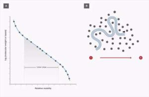
Figure 2. Migration of nucleic acids in gel electrophoresis. (A) Relationship between size and migration of linear double-stranded DNA fragments. (B) Biased reptation model.
A Brief History of Nucleic Acid Gel Electrophoresis
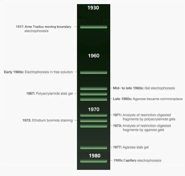
Electrophoretic separation of nucleic acids began with Tiselius’s pioneering work in 1937. He invented moving-boundary electrophoresis, laying the foundation for all modern electrophoresis techniques. In the early 1960s, nucleic acids were mainly separated by density-gradient centrifugation based on sedimentation velocity, with separation determined by nucleic acid size and conformation. Density-gradient centrifugation was not only time-consuming but also required heavy equipment and large sample amounts. Seeking a breakthrough, researchers began to explore the movement of DNA under an electric field in ionic or electrolyte solutions—that is, the process of electrophoresis.
Within a few years, by adapting techniques already used in protein electrophoresis, nucleic acid electrophoresis advanced further and began to use gel matrices as the separation medium. Through further exploration in the mid-to-late 1960s, agar (a natural carbohydrate), agarose (a component of agar), polyacrylamide (a synthetic gel), and agarose–acrylamide composite gels successfully became matrices for DNA and RNA electrophoresis. Early gel electrophoresis results showed correlations with sedimentation coefficients, or S values, from density-gradient centrifugation (the earlier established method for nucleic acid separation). With further understanding and development of agarose production in the late 1960s, agarose gradually replaced agar as the preferred gel electrophoresis medium. For practical powder selection today, see how agarose gel electrophoresis works and how to choose powder.
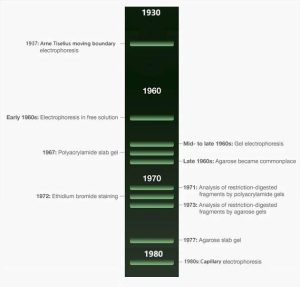
Figure 3. Timeline of early developments in nucleic acid gel electrophoresis.
In the 1970s, after the discovery of restriction endonucleases and their application in recombinant DNA technology, gel electrophoresis for separating and analyzing nucleic acids became more common. At that time, sucrose density-gradient centrifugation was the common separation method, but it was cumbersome and could not adequately distinguish similarly sized DNA fragments produced by restriction digestion. Because digestion of DNA from large to small fragments reduces solution viscosity, viscosity was empirically used as an indicator of successful restriction digestion. In 1971, Danna and Nathans first reported fractionation of restriction-digested SV40 DNA fragments by polyacrylamide gel electrophoresis, which revolutionized DNA fragment cloning. Although agarose and agarose–polyacrylamide gels had been used in the late 1960s to separate RNA and single-stranded DNA, studies analyzing restriction fragments by agarose gel electrophoresis were not published until around 1973 (Figure 3).
In the 1970s, major breakthroughs were also made in detecting nucleic acids after gel electrophoresis. Early electrophoresis methods relied on radioactive labeling of nucleic acids to visualize separated molecules. Although highly sensitive, radiolabeling was time-consuming and required radiation-safety training. In 1972, two laboratories independently described staining gels with the fluorescent molecule ethidium bromide (EtBr), which thereafter became a common, simple method for nucleic acid detection, with sensitivity down to a few nanograms of double-stranded DNA. Today’s fluorescent dyes are safer, more sensitive, and more specific than EtBr, further improving nucleic acid detection.
With the introduction of EtBr, and following the emergence of “slab” gels around 1970, gel electrophoresis became even more widely used and remains a cornerstone of today’s research and instrumentation. Early gel electrophoresis studies cast gels in glass tubes 1–3 mm in diameter. Throughput was extremely low because each tube could hold only one sample (Figure 4A). Vertical slab gels (Figure 4B) were simpler to prepare, allowed multiple samples to be run simultaneously, were first introduced for polyacrylamide in the late 1960s, and were further improved by researchers in the early 1970s. Horizontal agarose slab gels (Figure 4C), first introduced by McDonell et al. in 1977, are very similar to the format still used today. In the early 1980s, James W. Jorgenson and colleagues systematically proposed “capillary zone electrophoresis,” sparking a worldwide surge of research that ultimately led to the high-throughput capillary gel electrophoresis DNA sequencers that played a major role in the Human Genome Project. Today, mini precast, ready-to-use gels are available in both polyacrylamide and agarose for safer, faster, and simpler gel electrophoresis. In addition, some gel electrophoresis systems use precast bufferless gels that can be run in under 10 minutes. They can also be combined with digital imaging and analysis of separated nucleic acids for more efficient and convenient workflows.
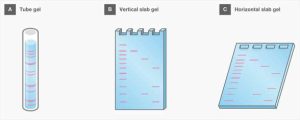
Figure 4. Common gel formats used in electrophoresis: tube gel, vertical slab gel, and horizontal slab gel.
In summary, gel electrophoresis has become a ubiquitous technique for separating nucleic acids in molecular biology. This analytical and preparative method is not only closely tied to common workflows such as molecular cloning and PCR, but also plays an important role in emerging technologies such as genome editing and next-generation sequencing. Labs that still cast gels from powder can source electrophoresis-grade Agarose (CAS 9012-36-6), review agarose applications and an introduction to agarose, or contact us for grades and pack sizes. Reagents and instruments for nucleic acid electrophoresis will continue to develop toward greener, more efficient, and more automated approaches, and—together with microfluidic chips and AI-based data analysis—will further expand applications in precision medicine and genetic diagnosis.