

Burn wound healing properties of asiaticoside and madecassoside

The healing of burn wounds has been widely characterized to be highly intricate, involving processes such as neovascularization, granulation, reepithelialization, inflammation, and wound contraction. Various therapies are available for the management of burn wounds; however, a truly effective therapeutic strategy has yet to be identified due to safety issues. The aim of the present study was to assess and confirm the burn wound healing properties of the compounds asiati-coside (AE) and madecassoside (MA), which are found in the herb Centella Asiatica. The cytotoxic nature of the AE and MA were inspected and were confirmed to be non-toxic up to 500 ppm. The compounds AE and MA increased monocyte chemoattractant protein-1 production but caused no significant effect on vascular endothelial growth factor production.
Despite the technological advancements of recent years, the utilization of herbal medicine as an alternative approach to the treatment of burn wounds remains limited. This is due to issues involving resistance, side effects, and the use of negative pressure wound devices, which affect the healing outcome. Among the widely available medicinal herbs, it has been previously reported that extracts from Centella Asiatica, Urban, which is also known as Asiatic Penny-wort, possesses properties able to cure certain injuries, including ulcerous skin abnormalities, burns, duodenal and stomach ulcers. In addition, C. asiatica extracts have been applied to the treatment of lupus, scleroderma, leprosy, and diseases of the veins. The mechanisms of action of this extract are speculated to include the synthesis of collagen and stimulation of extracellular matrix accumulation. There are numerous active compounds in C. asiatica, including several triterpenoid saponins; madecas-soside (MA), asiatic acid, and asiaticoside (AE) .
A previous study investigated the efficacy of using the active compound trisaccharide triterpene in wound healing and demonstrated that it was able to increase the levels of anti-oxidants in a rat model of initial-stage excision-type cutaneous wounds. Furthermore, it has been reported that procollagen mRNA types I and III isoforms and their protein production, as well as the proliferation of human dermal fibroblasts, are increased with exposure to C. asiatica. Alternatively, C. asiatica has been utilized as a complementary medicine that can be administered orally or topically to stimulate wound recovery.
Furthermore, the compound MA, which is found in C. asiatica, has been reported to stimulate the expression of collagen and is speculated to possess immunomodulatory properties. Notably, Haftek et al conducted a double-blind, randomized clinical investigation, which indicated substantial improvement in the clinical score for suppleness, firmness, wrinkles, roughness as well as skin hydration with the use of C. asiatica.
In addition, when the extract is ingested orally, the active compounds present have been reported to improve the regeneration of nerve tissue apart from stimulating neurite elongation in an in vitro setting. Further to this, the extract has similarly demonstrated the ability to increase neurite formation, particularly in the SH-SY5Y human cell line with the addition of neuronal factors. A predominant factor involved in functional recovery following burn wounds is axonal regeneration, and this was also noted with the presence of non-polar fraction. Normally, in cases of burn injuries, the first line of defense includes a well-orchestrated, complex wound-healing process. As a result of the directly afflicted burn injury, the central area of coagulation is comprised of tissue as well as cell necrosis. Monocyte-derived macrophages and neutrophils are then attracted to the site as a response to the selection of cytokines and chemoattractants released by platelets in the hemostatic clot. Furthermore, it should be noted that burn wound healing requires a series of intricate phases which include neovascularization, re-epithelialization, inflammation, granulation, neovascularization, and wound contraction. Throughout this process, a selection of biomessengers is thus activated

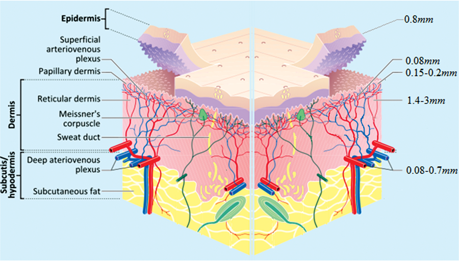
including, cytokines, liver and kidney damage biomarkers, and antioxidants. Burn wounds are normally characterized as tissue injury due to external causes such as chemicals, radiation, heat, and electricity. As a result, the outcome of burn wounds generated may be further grouped into superficial (first degree), partial thickness (second degree), or full-thickness (third degree), primarily based on the depth of the wound. Wound healing following burns involves a multitude of processes, including inflammation that disrupts blood vessels and induces ̔leakage̓ of blood constituents into the target area which then stimulate re-epithelialization. Subsequently, granulation tissue comprised of macrophages and fibroblasts is formed and this complex tissue is responsible for extracellular matrix recovery, and neo-vascularization towards the target area in addition to the mitogenic stimulation and migration of cells of endothelial origin. Finally, contraction of the wound occurs as a result of communication between extra-cellular matrix cells as well as cytokines. Due to the potential healing properties of medicinal plants, alkaloids such as flavonoids, essential oils, fatty acids, tannins, saponins, terpenoids, and phenolic compounds, may be useful in improving the healing process. Furthermore, medicinal plants are renowned for their availability, cost effectiveness and an association with fewer side effects compared with FDA approved drugs. Thus, substantial interest exists globally, concerning the identification and isolation of the active components from therapeutic plants which may promote wound healing.
Studies have shown, it was revealed that AE and MA possess wound healing properties and are able to stimulate collagen synthesis, reducing wound oxidative stress as well as inducing vasodilatation. It was also indicated that AE and MA have positive effects on proliferation and cell growth in animal models. Previous studies have reported that these compounds may restore tissue functionality via activation of growth factors, including VEGF, endothelial growth factor and fibroblast growth factor.