

Nucleic acid electrophoresis is used in many molecular biology research applications to verify experimental results, and sometimes to separate and purify samples for downstream use. Conventional applications are therefore often grouped into analytical and preparative types; both rely on separation, resolution, and quantification. These applications include:
1. Analytical electrophoresis applications
- PCR, restriction digestion, ligation, colony screening, reverse transcription, in vitro transcription
- Gel quantification
- Nucleic acid purity, sample fragmentation, oligonucleotide synthesis efficiency
- Southern blotting, Northern blotting, nuclease protection assays
- DNA conformation, EMSA
2. Preparative electrophoresis applications
- PCR, restriction digestion, oligonucleotide synthesis, NGS size selection
- Agarose gel purification
1. Analytical electrophoresis for confirming experimental results
Before moving to the next workflow step or another set of experiments, analytical nucleic acid electrophoresis can be used to check experimental outcomes. As described below, this approach mainly evaluates whether the target band is present in the gel, as well as band intensity, migration pattern, mobility, and hybridization.
1a. Checking the success of enzymatic synthesis, digestion, and cloning experiments
Electrophoretic analysis of nucleic acids is commonly performed immediately after the following techniques (Figure 1) to determine experimental success and efficiency:
- Within a few hours, polymerase chain reaction (PCR) amplifies one copy of a target sequence into millions of copies. Electrophoresis after endpoint PCR confirms target amplification and yield.
- After restriction digestion with a restriction enzyme that cuts specific sequences on a DNA substrate, samples are run on a gel to determine the DNA cutting pattern (and how complete the digestion is).
- In molecular cloning, DNA fragments are inserted into a vector by ligation. In some cases, electrophoresis can be performed after ligation to assess reaction efficiency (Figure 2). Ligation products are then used to transform competent cells of a cloning host such as E. coli for propagation, followed by colony screening to determine whether colonies carry a vector with the desired insert. PCR and restriction digestion—both of which commonly include electrophoresis as part of the workflow—are frequently used in colony screening.
- Reverse transcription synthesizes DNA complementary to an RNA template (cDNA) using reverse transcriptase. After cDNA synthesis and removal of the RNA template, products can be run on a denaturing gel to assess reaction efficiency. This approach is most common when reverse-transcribing RNA of known sequence or known length.
- In vitro transcription synthesizes RNA from a DNA template using RNA polymerase. After removal of the DNA template, the synthesized RNA can be electrophoresed on a denaturing gel to confirm successful transcription.
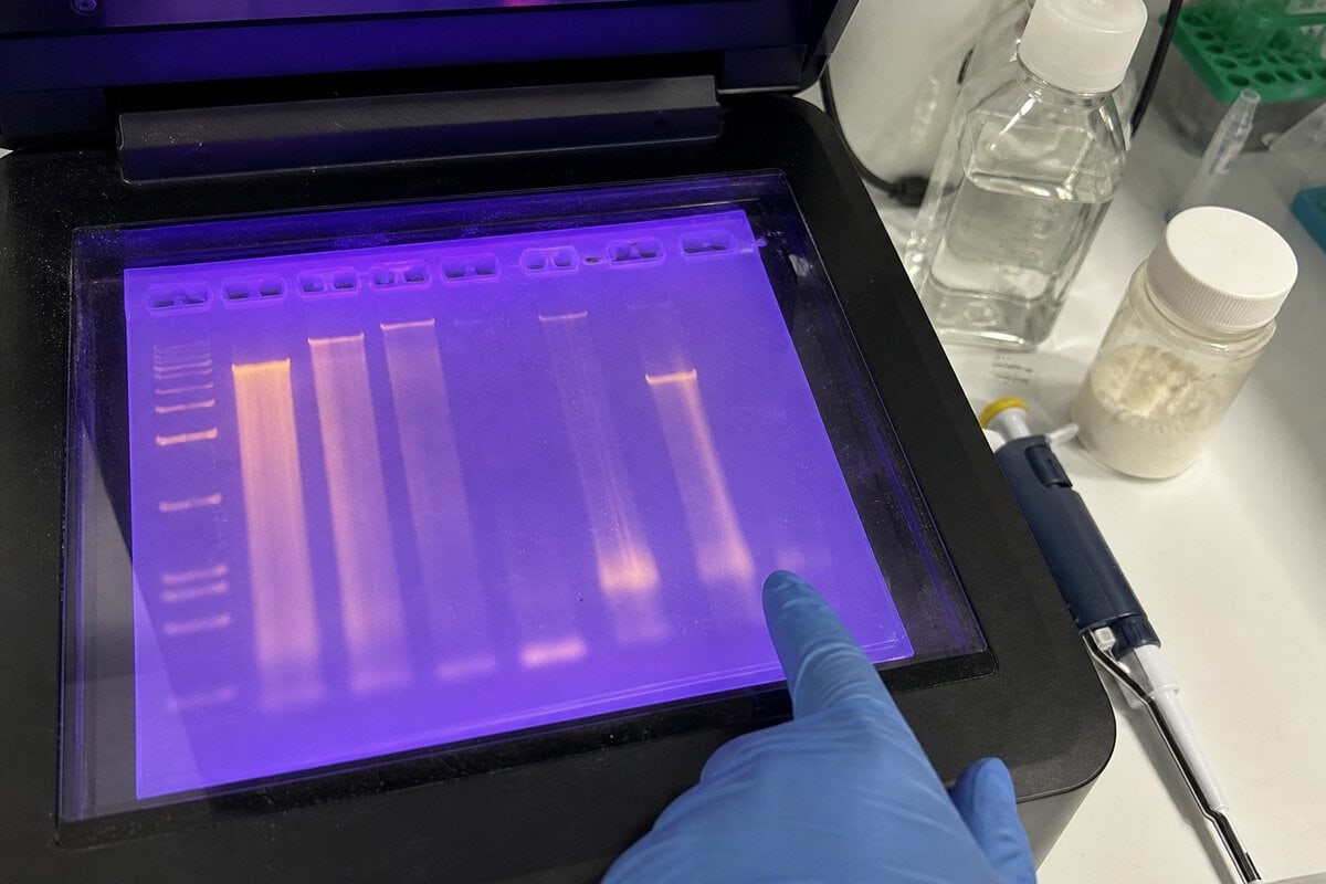

Figure 1. Common molecular biology applications in which electrophoresis is used to confirm experimental success.

Figure 2. Determining ligation efficiency by gel electrophoresis. λ DNA was first cut with HindIII, a Type II site-specific restriction endonuclease (lane 1). The sample was then religated and analyzed by gel electrophoresis (lane 2); fully ligated DNA appears as the most prominent band.
1b. Quantifying nucleic acid samples by gel electrophoresis
Electrophoresis can quantify a DNA or RNA band of interest using standards or ladders in which the amount of each fragment is known. Common spectrophotometric quantification can be affected by contaminants such as nucleotides and primers. Electrophoresis, however, separates the target sample from these contaminants and is therefore a reliable quantification method.
Gel quantification estimates sample amount by comparing the intensity of a band in the sample with a similarly sized band in the ladder (Figure 3A). Ideally, when using a ladder designed for quantification, different amounts of ladder are loaded to plot a standard curve of amount versus intensity for more precise quantification (Figure 3B). Staining nucleic acids with fluorescent dyes that have high sensitivity and a wide dynamic range can further improve gel quantification. Some gel imagers include analysis software for simpler quantification of samples in the gel, while others offer cloud connectivity for data storage.

Figure 3. (A) Gel quantification using fragments of known amount. (B) Standard curve for gel quantification.
1c. Analyzing sample purity, integrity, fragmentation, and synthesis efficiency
Gel electrophoresis can assess the purity and integrity of nucleic acid samples extracted from their source, the success of sample fragmentation, and the percentage of full-length oligonucleotide after synthesis.
- Genomic DNA contaminating an RNA sample—or vice versa—can be detected by gel electrophoresis to assess sample purity (Figure 4A). Detection of contaminating species depends on the sensitivity of the nucleic acid stain used and the amount of contaminant.
- Using gel electrophoresis, the integrity of total RNA after extraction can be checked by evaluating the relative intensities of 28S and 18S rRNA, where a 2:1 ratio indicates intact RNA. Smearing, especially at lower molecular weights, indicates RNA degradation (Figure 4B).
- Some protocols require fragmentation of nucleic acid samples—for example when preparing input for chromatin immunoprecipitation (ChIP) and next-generation sequencing (NGS)—to obtain fragment sizes suitable for the next step. Fragmentation efficiency can be checked by gel electrophoresis (Figure 4C).
- After synthesis, because coupling efficiency is <100%, oligonucleotide products contain full-length as well as truncated or failed sequences (i.e., n−1, n−2, etc.). Electrophoresis can distinguish full-length products from failure sequences based on the size and conformation of oligonucleotides of different lengths.

Figure 4. Assessing sample purity, integrity, and fragmentation by gel electrophoresis. (A) Extracted genomic DNA and total RNA analyzed on separate gels. Red arrows indicate contaminating RNA and genomic DNA, respectively. Contaminating RNA is detectable only at the start of the run (≤5 minutes). (B) Integrity of two purified RNA samples assessed by electrophoresis and analysis of 28S and 18S rRNA. (C) Efficiency of DNA fragmentation and the distribution of fragmented DNA measured on a gel. (M = molecular-weight marker. RNA samples were run on denaturing gels.)
1d. Detecting target sequences in mixed samples
Gel electrophoresis is a key part of workflows that detect target sequences in nucleic acid libraries by probe hybridization (Figures 5 and 6). A probe is a single-stranded nucleic acid of known sequence designed to bind the target sequence by base complementarity.
- For DNA fragment analysis, Southern blotting and restriction fragment length polymorphism (RFLP) analysis are two well-known techniques in which electrophoresis separates samples before target detection (Figure 5).
- For RNA fragment analysis, Northern blotting and nuclease protection assays (NPA) also rely on probe hybridization to detect sequences of interest. Aside from the input being RNA (Figure 5), Northern blotting follows the same workflow as Southern blotting. In an RNase protection assay (RPA), an RNA probe binds the target sequence in the sample mixture. Remaining single-stranded RNA—such as unbound probe and template and their overhangs—is then digested by RNases such as an RNase A/T1 mix. Bound probe and sample are subjected to gel electrophoresis for downstream target detection and analysis.

Figure 5. Detection of specific DNA and RNA sequences in mixed samples using Southern and Northern blotting, respectively

Figure 6. Nuclease protection assay.
1e. Assessing DNA conformation and nucleic acid–protein complexes
As discussed in the electrophoresis section, plasmid DNA of the same sequence but different conformations can show different electrophoretic mobilities. This feature can be used to assess DNA conformation and the level of intact plasmid after extraction.
Nucleic acid fragments bound to protein migrate more slowly than unbound fragments. Methods based on this principle are formally called electrophoretic mobility shift assay (EMSA), and are often referred to as gel-shift or gel-retardation assays because the bound protein shifts or retards migration of the nucleic acid fragment on the gel (Figure 7). The electrophoresis step of the experiment can therefore “capture” the equilibrium between bound and free DNA in the sample. For EMSA, low-ionic-strength buffers should be used in gel preparation and electrophoresis to help stabilize nucleic acid–protein complexes during the run.

Figure 7. Electrophoretic mobility shift assay (EMSA).
2. Preparative electrophoresis for purifying nucleic acid samples
Preparative applications of nucleic acid gel electrophoresis refer to purifying and extracting separated nucleic acids from the gel matrix after electrophoretic analysis. Electrophoresis used for gel purification is therefore often a preparatory step for downstream applications. In addition, preparative gel methods can be combined with analytical methods.
2a. Applications related to preparative electrophoresis
Preparative electrophoresis is commonly used after many molecular biology techniques and operations, most often: PCR; restriction digestion; oligonucleotide synthesis; and the fragmentation, end-modification, and ligation steps of next-generation sequencing (NGS).
- PCR products and restriction-digested DNA can be purified from gels for downstream applications such as end modification, ligation, cloning, and sequencing.
- After oligonucleotide synthesis, polyacrylamide gel electrophoresis is one of the main methods for separating and purifying full-length oligonucleotides from salts and truncated sequences.
- For Illumina and Ion Torrent NGS platforms, as part of sequencing library preparation, samples are fragmented, end-modified, and ligated to adapters. After these steps, DNA fragments in a specific size range (e.g., 200–300 bp) are purified by a method called size selection—not only to remove low- and high-molecular-weight fragments, but also adapters, enzymes, and reagents from previous steps. Size selection helps ensure that input samples have fragments of uniform length, enabling high-quality, consistent sequencing [1]. Gel electrophoresis is one size-selection method because of its high efficiency in selecting fragments of a specific size within a narrow range (Figure 8).

Figure 8. Fragment size distribution before and after size selection.
2b. Overview of gel purification
Preparative electrophoresis is a key step in separating and purifying nucleic acids from gels. Nucleic acid purification usually starts by excising the target sample band from the gel matrix, then extracting the nucleic acid. When performing gel purification, note the following:
- Minimize the gel area excised to facilitate extraction.
- Prevent damage to nucleic acids during gel visualization.
If using a UV-excitable dye (e.g., ethidium bromide) for visualization, minimize sample exposure to radiation, use a longer UV wavelength for excitation (e.g., 360 nm rather than 254 nm), and prefer epi-illumination over transillumination. Fluorescent dyes that can be excited by lower-energy blue light (e.g., SYBR dyes) are better options for sample visualization because they reduce radiation damage to the sample.
Methods for purification from agarose gel matrices are summarized below:
After cutting out the target band, agarose can be melted by heating. At 1% gel concentration, standard agarose melts at >90°C, whereas low-melting-point (LMP) agarose melts at >65°C. Because of its lower melting point, LMP agarose can improve integrity and yield of extracted nucleic acids. For a gentler method than heating, consider enzymatic agarase, which breaks agarose chains into oligosaccharides. Agarase digestion requires LMP agarose so that agarase remains active at the low gelling temperature of LMP agarose.
After melting or digesting the agarose, nucleic acids can be extracted by any of the following methods. One simple and effective approach is phenol extraction combined with ethanol precipitation. Because of phenol’s toxicity and the need to dispose of it as organic waste, a popular current method is to bind nucleic acids to a silica-based spin column in the presence of a chaotropic agent, then elute them from the column (Figure 9).

Figure 9. Extraction of DNA from a gel using a silica column.
In summary, nucleic acid gel electrophoresis is widely used across molecular biology workflows and techniques. Although the basic method of electrophoresis has changed little since the 1970s, electrophoresis has become an established, powerful technique for nucleic acid separation and analysis in applications ranging from restriction digestion to next-generation sequencing.